|


|
6. Chemical and biological
crystallography
6.1 Laboratory of
Chemical and Biological Crystallography, Rudjer Bošković Institute
Biserka
Kojić-Prodić
LCBC 2008
The X-ray structure analysis
in Croatia was initiated by Drago Grdenić after his return from his
doctoral studies carried under guidance of professors A. N. Nesmejanov
and A. I. Kitaigorodski in the Institute of Organic Chemistry of
Academy of Science in Moscow (1946-1948). His first rotation photograph
was taken by Unicam S-25 camera on New Year's Eve 1948 in the Physics
Department of the Faculty of Science where professor Mladen Paić already
had set up X-ray diffraction equipment for powder diffraction. The
results obtained in Zagreb were presented in three communications in
1949, 1950, and 1952. In 1950 by efforts of Boris Kidrič (politician),
and Ivan Supek ( physicist), the Federal Goverment Decree guaranteed
the foundation of the institute for fundamental research in physics
and chemistry in Zagreb. The interdisciplinary Rudjer Bošković Institute
founded on the concepts of professor Ivan Supek enabled the research to
scientists from the University of Zagreb who joined new young
researchers of the Institute in developing science in Croatia. In 1951
the first Weissenberg goniometer was purchased together with Ilford
films required for recording diffraction intensities, and subscription
to Acta Crystallograpica was provided. In those days, facilities
for X-ray structure analysis in Zagreb were not different from those in
Europe. Professor Drago Grdenić was the first head of The Department of
Structural and Inorganic Chemistry, constituted upon his concepts and
founded in 1952, including X-ray Laboratory. The most significant
scientific results from that period were determinations of crystal
structures of organic molecules, phthalylurea (published in 1953) and
mellitic acid (published in Nature, 1960) solved by original
direct methods designed by Aleksadar Bezjak under supervision of D.
Grdenić. In those days neither sophisticated computers nor computer
programs were available and structures were
solved from projections. The existence of a novel mercurated
oxonium species, as a part of PhD thesis of Stjepan Šćavničar under
supervision of D. Grdenić, was published in Nature (1953).
The structure of thorium(IV)acetylacetonate, solved by
Boris Matković under supervision of D. Grdenić, revealed an Archimedean
antiprism as the coordination polyhedron around thorium. For the first
time such geometry was detected for an octa-coordination and the result
was published as a short communication in Nature (1958).
In early fifties of 20th century, the X-ray laboratory was set up on the
experiences from British, French, and Russian crystallographic schools.
From the first days of the
foundation of the X-ray Laboratory to the present days two research
lines have been
maintained: to develop and implement experimental
techniques and
 numerical
methods needed in the X-ray structure analysis. During the forthcoming
years the research was focused on synthesis of heavy metal alkaline
phosphates by a melt technique and determination of their structures
from projections; among them a new type of ferroelectric substance
without hydrogen bond was discovered. Later on, the interest was shifted
to transition metal complexes and their structures. At the time when
direct method programs, such as Multan, were available, structures and
conformations of organic molecules, including pharmaceutically and
biologically active molecules, were the objects of research. In addition
to studies of various groups of organic compounds the research involved
plant growing hormone auxin and its analogues, peptides, sugars, and
pharmaceutically active compounds (cimetidine, ranidine, pyroxiam,
diltiazem and some of their derivatives). In search on
structure-activity correlations the spectroscopic methods, bio-assays,
and molecular modelling were used. Since 1985 Camridge Structural
Database has been available in Croatia via National Affiliation Centre
of Cambridge Crystallografic Data Centre. In 1989 a first
diffractometer with a point detector was purchased. Over
forty years the research was dedicated to chemical crystallography in
order to provide data on structural characteristics and properties of
molecules, to understand intramolecular and intermolecular interactions,
and to visualise the crystal packing and topology of hydrogen bonds. As
the activities were more and more concentrated on biologically active
molecules and their interactions, the Laboratory for Chemical and
Biological Crystallography was founded in 1997 with the aim to introduce
protein crystallography. Since then interactions of enzymes
(hydrolases) with substrates and inhibitors have been studied in order
to resolve their catalytic mechanisms at molecular level. The study of
proteins and their interactions relevant for a cell life has been active
since 2006. Progress in protein crystallography has been
supported by in-house modern diffractometer installed at the end of 2007
and by the efforts of the team to introduce biochemical and genetic
methods. numerical
methods needed in the X-ray structure analysis. During the forthcoming
years the research was focused on synthesis of heavy metal alkaline
phosphates by a melt technique and determination of their structures
from projections; among them a new type of ferroelectric substance
without hydrogen bond was discovered. Later on, the interest was shifted
to transition metal complexes and their structures. At the time when
direct method programs, such as Multan, were available, structures and
conformations of organic molecules, including pharmaceutically and
biologically active molecules, were the objects of research. In addition
to studies of various groups of organic compounds the research involved
plant growing hormone auxin and its analogues, peptides, sugars, and
pharmaceutically active compounds (cimetidine, ranidine, pyroxiam,
diltiazem and some of their derivatives). In search on
structure-activity correlations the spectroscopic methods, bio-assays,
and molecular modelling were used. Since 1985 Camridge Structural
Database has been available in Croatia via National Affiliation Centre
of Cambridge Crystallografic Data Centre. In 1989 a first
diffractometer with a point detector was purchased. Over
forty years the research was dedicated to chemical crystallography in
order to provide data on structural characteristics and properties of
molecules, to understand intramolecular and intermolecular interactions,
and to visualise the crystal packing and topology of hydrogen bonds. As
the activities were more and more concentrated on biologically active
molecules and their interactions, the Laboratory for Chemical and
Biological Crystallography was founded in 1997 with the aim to introduce
protein crystallography. Since then interactions of enzymes
(hydrolases) with substrates and inhibitors have been studied in order
to resolve their catalytic mechanisms at molecular level. The study of
proteins and their interactions relevant for a cell life has been active
since 2006. Progress in protein crystallography has been
supported by in-house modern diffractometer installed at the end of 2007
and by the efforts of the team to introduce biochemical and genetic
methods.
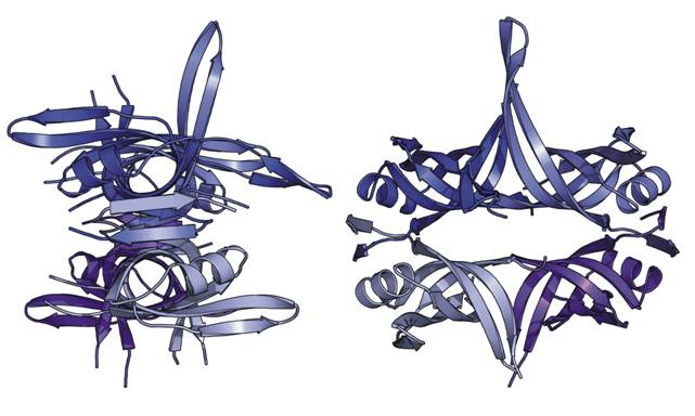
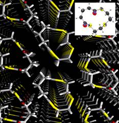
Two views of the single-stranded DNA-binding protein from
Streptomyces coelicolor, the first X-ray structure of an SSB protein
from a member of the genus Streptomyces (Z. Štefanić, D.
Vujaklija and M. Luić, Acta Cryst. (2009). D65, 974–979).
6.2 Powder diffraction,
Ruđer Bošković Institute
Stanko
Popović
In the X-ray Laboratory,
powder diffraction was also used for the phase analysis and studies of
micro and nano crystalline materials. New methods for accurate
unit-cell parameter measurement were proposed: 1st, which was based on
the separation of adjacent diffraction lines rather than on their
angular positions (J.Appl. Cryst.6(1973)122,411);
2nd, which combined single-crystal zero-layer rotation and Weissenberg
patterns with powder diffraction pattern (J.Appl. Cryst.7(1974)291).
The methods for quantitative phase analysis were introduced, in which a
multicomponent sample was doped with those components the fractions of
which were to be determined ( J.Appl.Cryst. 12(1979)205;
16(1983)505 ). Phase diagrams of semiconductors (Ax B1-x
)2III(Cx D1-x ) 3VI
and AI(BxC1-x)IIID2VI
were systematically studied and related to electrical properties (e.g.
J.Appl.Cryst.13(1980)24). High temperature phases of In2Se3
were defined (J.Appl.Cryst. 12(1979)416). Microstructure
and phase diagrams of a series of mixed metal oxides (e.g. ferrites,
orthoferrites, alumina, zirconia, titania), prepared along different
procedures, were systematically studied in the whole concentration
region by X-ray powder diffraction, Raman, FT-IR and Moessbauer
spectroscopies, in collaboration with the Department of Materials
Chemistry, Rudjer Bošković Institute. The processes of biomineralization
of bivalvia have been studied in detail in collaboration with the
Center for Marine Research, Rudjer Bošković Institute (Marine Biology
129(1997)615).
|